
The June 2026 issue of the Research Resource Nexus highlights the Magnetic Resonance Research Center (MRRC), a state-of-the-art facility located on the eighth floor of UPMC Presbyterian dedicated to magnetic resonance imaging (MRI) for medical and biological research. MRRC provides whole-body imaging capabilities with specialties in anatomical MRI imaging of the brain, spine, liver, bladder, joints, muscles and more. They also offer functional MRI to study cognitive, sensory and motor function in the brain.
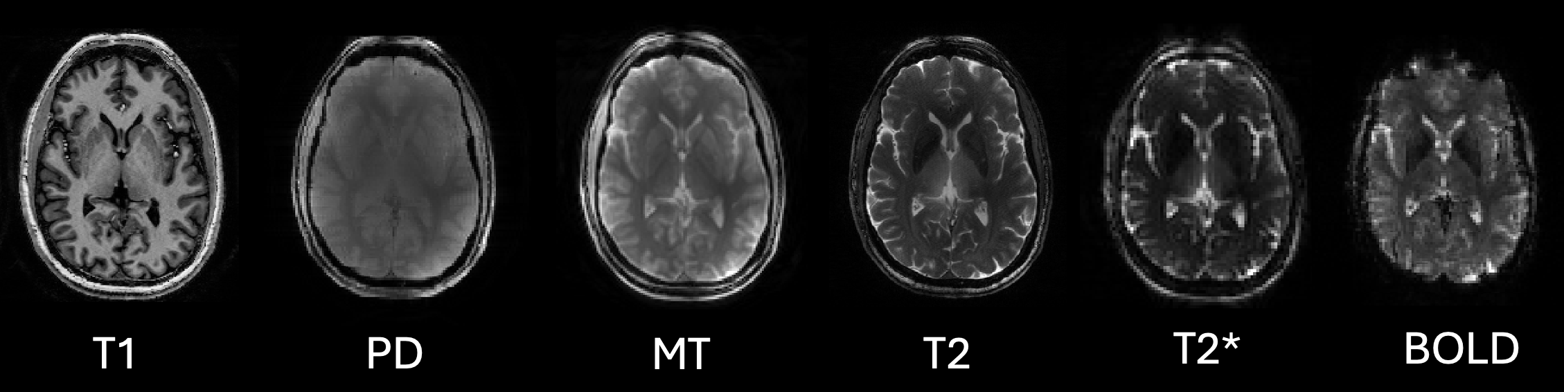
The facility contains three 3-Tesla (3T) high-field MRI systems for standard research-grade imaging and a brand new 7-Tesla (7T) Siemens TerraX MRI for high-resolution imaging and to answer research questions that require ultrahigh field strength. For context, the typical MRI in a clinical setting is a 1.5T system, and a 3T system is 45,000-100,000 times stronger than Earth’s magnetic field. Approximately 80% of imaging at MRRC involves the brain, supporting both basic and clinical investigators largely in neurology, neurosurgery, psychiatry and psychology disciplines.
MRRC technologies include, but are not limited to, the following, with industry-standard MRI acronyms in parentheses:
- anatomical/structural scans (T1, T2 and FLAIR),
- cerebral blood flow (ASL and pCASL),
- brain chemistry and metabolite measurement (MRS and MRSI),
- functional activity tracking (BOLD/VASO fMRI),
- white-matter pathway reconstruction (DTI and RESOLVE),
- dopamine-producing neurons visualization (neuromelanin imaging),
- high-order shimming for clean signal at ultra-high field, and
- data processing and analytics support.
Getting Started With MRRC
Investigators must contact MRRC staff prior to grant submission to discuss imaging needs and ensure the center can support the study appropriately. Once funding is acquired, MRRC faculty and MRI technologists will work with the investigator’s team to develop a study protocol. This includes an in-depth onboarding process to build and refine the appropriate imaging protocol on the scanner to validate details such as image location, resolution and sequence parameters. The study team, which includes any individual who wants access to restricted areas of the MRRC (e.g., primary investigators, coordinators, research assistants), is required to complete MRI safety training. MRRC follows multiple levels of rigorous training to ensure MRI safety for staff, study teams and study participants.
With 80-100 active projects in the last 90 days and more than 400 hours of scanning per month, MRRC has become one of the most dominant brain imaging centers and the gold standard for high-resolution, high-field human imaging on Pitt’s campus.
To initiate a project or get more information, contact MRRC Research Operations Manager Andrew Reineberg at aer126@pitt.edu or visit the MRRC website.
Testimonials
Keith Vogt, associate professor of anesthesiology and perioperative medicine, School of Medicine, and a physician-anesthesiologist, has used MRRC for more than a decade through his Anesthesiology Neuroscience Research Program to study how different anesthetics act in the human brain to suppress pain and memory. In this research, functional MRI is not just a tool; it is the scientific centerpiece. Whole-brain imaging is the only approach that can simultaneously reveal drug-induced changes in neural activity and network architecture with the spatial resolution needed to distinguish the deep brain regions central to pain experience and memory formation. MRRC's location within UPMC Presbyterian enabled the use of intravenous and inhaled anesthetics across Vogt’s studies by providing access to medical gases and immediate emergency clinical support. The center's robust array of nearly identical 3T scanners dedicated to research has facilitated study throughput, especially considering the complex setup and close anesthesia monitoring his studies require. Vogt’s research, supported by the National Institute of General Medical Sciences, National Institutes of Health, plans to leverage additional MRRC resources in the future, including the electroencephalography acquisition system and newly installed 7T scanner.
Adriane Soehner, associate professor of psychiatry, School of Medicine, and director of the Sleep, Affective Neuroscience and Development Lab, is investigating neuro-affective responses to light among adolescents and adults with symptoms of depression through her DAYLight study. Morning bright light therapy is a low-cost, low-burden evidence-based treatment approach for depression, but it remains unclear how it works and whether its clinical benefits vary by age. To answer these questions, MRRC worked with Soehner’s team to develop an optimized scanning protocol using an MRI-compatible fiber optic cable and a custom 3D-printed mount to deliver targeted light stimuli to participants during MRI scans. This enabled precise light stimuli at a specific distance from the cornea with concurrent eye tracking to understand how light affects the brain. Soehner noted that the MRRC team provided solutions ranging from highly practical to deeply technical to make this protocol happen.