
Regenerative medicine research aims to restore the structure and function of damaged tissues and organs using approaches such as tissue engineering, cellular therapies and medical devices. This edition of the Research Resource Nexus highlights the McGowan Institute Histology Core Laboratory, one of the histology cores available within the University of Pittsburgh Health Sciences, located in the McGowan Institute for Regenerative Medicine (MIRM) at Bridgeside Point II. While primarily serving MIRM investigators, the core supports researchers across the Pitt community by providing specialized expertise to help them better understand the complex interactions between native tissue and engineered materials, and offering a full range of histology services.
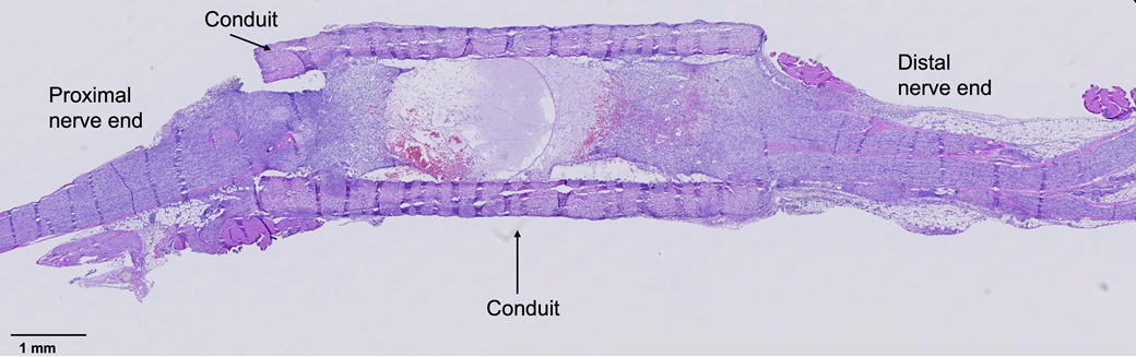
Capabilities include
- tissue processing and embedding (paraffin and cryo),
- sectioning, including multilevel and multispecimen slides,
- routine and special stains (e.g., H&E, Toluidine Blue and Trichrome),
- immunohistochemistry staining and antibody workup,
- whole slide imaging, and
- sample preparation for spatial biology workflows (e.g., Xenium and GeoMx).
In addition to these services, the McGowan Histology Core works closely with investigators to tailor workflows to the specific needs of each project. This is especially important for regenerative medicine applications, where samples often include embedded materials, such as polymers or medical devices. Core staff frequently consult with researchers early in the experimental design process to advise on tissue preparation, embedding strategies and sectioning approaches that will yield optimal results. When needed, investigators can work alongside core staff during embedding to ensure proper orientation of nontraditional samples.
The core also offers flexible options for sectioning and analysis, including multilevel cutting to visualize tissue at different depths and the ability to mount multiple specimens on a single slide for comparative studies. For larger or more complex specimens, the core provides “Supa Mega” large format sectioning, which uses slides approximately twice the size of standard formats, to preserve larger tissue samples and eliminate the need for image stitching. For spatial transcriptomics workflows, the core supports specialized preparation for Xenium and GeoMx platforms, including maintaining strict RNase-free conditions and coordinating sample handoff to preserve integrity.
Researchers can initiate a request through iLab, or contact McGowan Histology Core Supervisor, Julia Hart, at jrp128@pitt.edu with questions about services, specialized projects or staining requests. Information about transportation, parking and sample drop-off is available on the core’s iLab site.
Testimonials
“The Histology Core at the McGowan Institute for Regenerative Medicine is far more than a technical service facility—it is a strategic research partner that dramatically accelerates scientific discovery. Their exceptional expertise in tissue preparation for spatial transcriptomics and spatial proteomics enables investigators to generate high-quality datasets on the first attempt, minimizing costly delays and experimental failures. Laboratories that leverage the core gain immediate access to world-class histological expertise, allowing trainees and researchers to focus their time on data interpretation and scientific innovation rather than troubleshooting tissue preparation. The resulting gains in productivity, efficiency and data quality can substantially increase the pace and impact of a laboratory’s research program.”
—Chandan K. Sen, director, McGowan Institute for Regenerative Medicine
“Unlike routine diagnostic histology—which processes soft tissues like biopsies or organ samples using standard paraffin embedding—biomaterial histology must evaluate the complex, intimate interface between living host tissue and foreign engineering materials. The MIRM Histology Core is unique in that it handles diverse, nonroutine materials like hydrogels, polymer scaffolds, calcified or soft engineered constructs while tailoring tissue processing, embedding and staining protocols to meet specific, complex research needs. Whether it’s evaluating the foreign body response, inflammation, fibrous capsule thickness and direct cell-to-material attachment, the expert histopathologists of the MIRM core provide timely, high-quality analysis of how living tissues interact with engineered implants, scaffolds or grafts.”
—George S. Hussey, scientific director, MIRM Histology Core
Acknowledge Core Facilities in Your Publications
Proper acknowledgment helps cores secure continued funding and recognition.
Please cite the featured core as follows:
Work performed in the McGowan Institute Histology Core Facility (RRID:SCR_027956) and services and instruments used in this project were graciously supported, in part, by the University of Pittsburgh and the McGowan Institute for Regenerative Medicine.
Find citation guidelines for all Pitt Health Sciences core facilities.